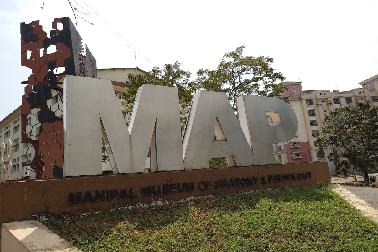
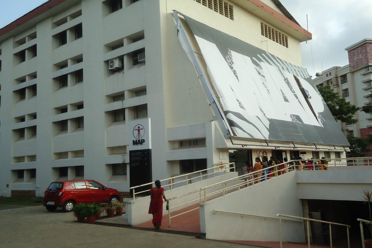
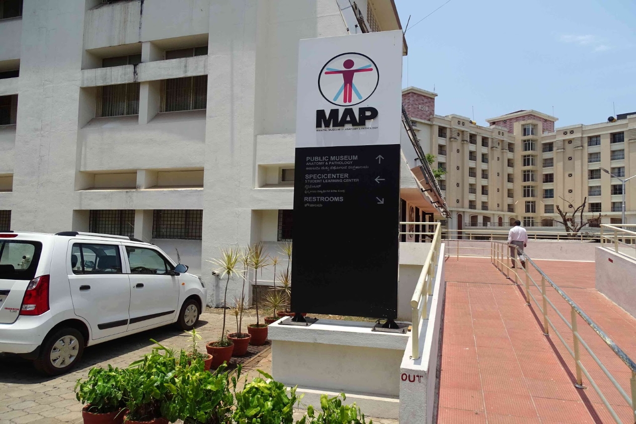
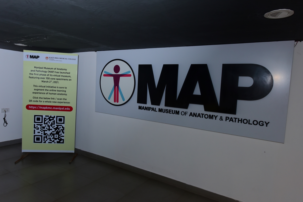
Manipal Museum of Anatomy and Pathology (MAP)
On most days, there is a serpentine queue outside the Manipal Museum of Anatomy and Pathology, or MAP, a sprawling building diagonally opposite the main education building.
For students, it is a wonderful learning aid and for the visitors, MAP is a storehouse of knowledge. The specimens have been there for close to 40 years. The renovated museum with its new magnificent ambiance will be an added attraction for hundreds and thousands of visitors.
Apart from college and school students, some from as far away as Mysore and Mumbai, there is a usually scattering of medical professionals and tourists from around the world, jostling to get in.
Timings- 9.00 AM-5.30 PM
(Tickets will be issued only until 5pm)
November, December, and January: Open on all days
February to October: Closed on Tuesdays
Closed on Public Holidays: Independence Day, Ganesh Chaturthi, and Deepavali
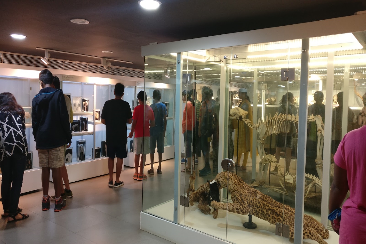
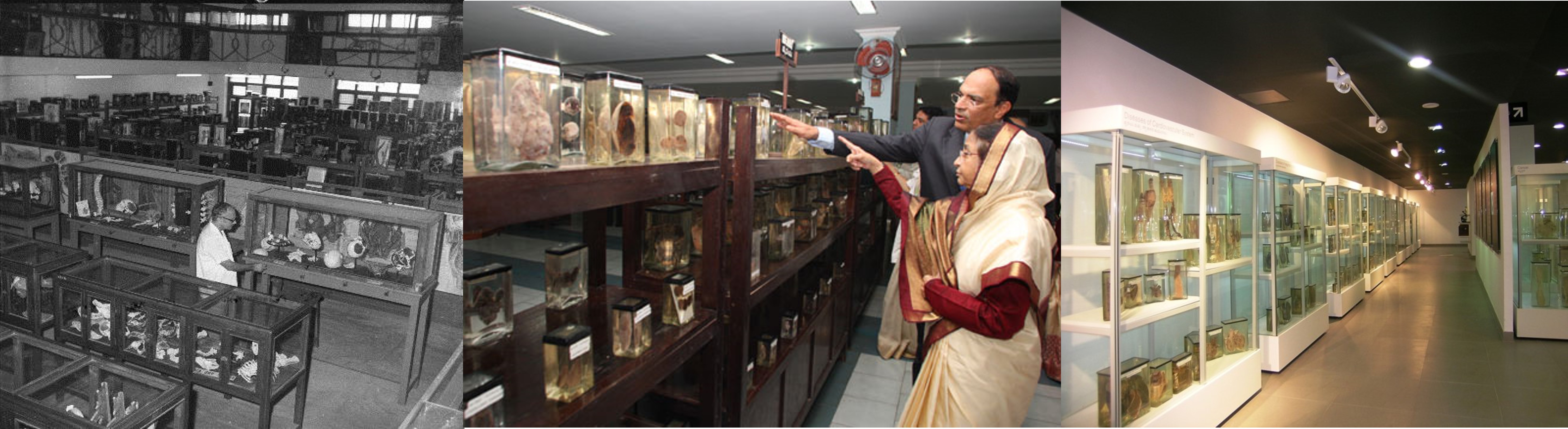
Billed as one of the largest in Asia, the museum boasts of over 3,000 specimens and samples of things anatomical, including the skulls of an elephant and a whale, and the long skeleton of a King Cobra.
Dr. SS Godbole, the first Anatomy Professor of Kasturba Medical College, had a passion for preservation. His techniques of careful dissection, processing, and mounting anatomical specimens are followed even today. It was under his watch that the museum opened its doors to the public in 1954, with over 650 specimens from his collection.
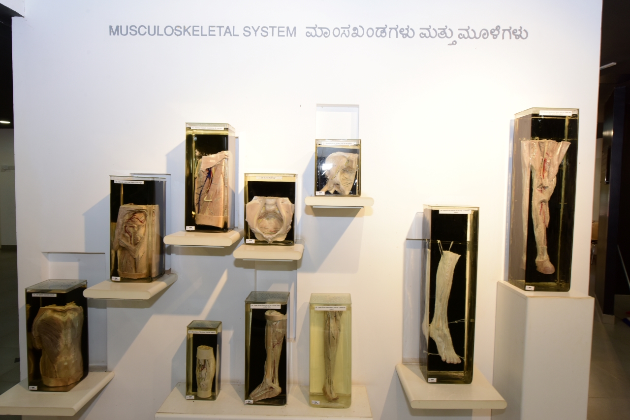
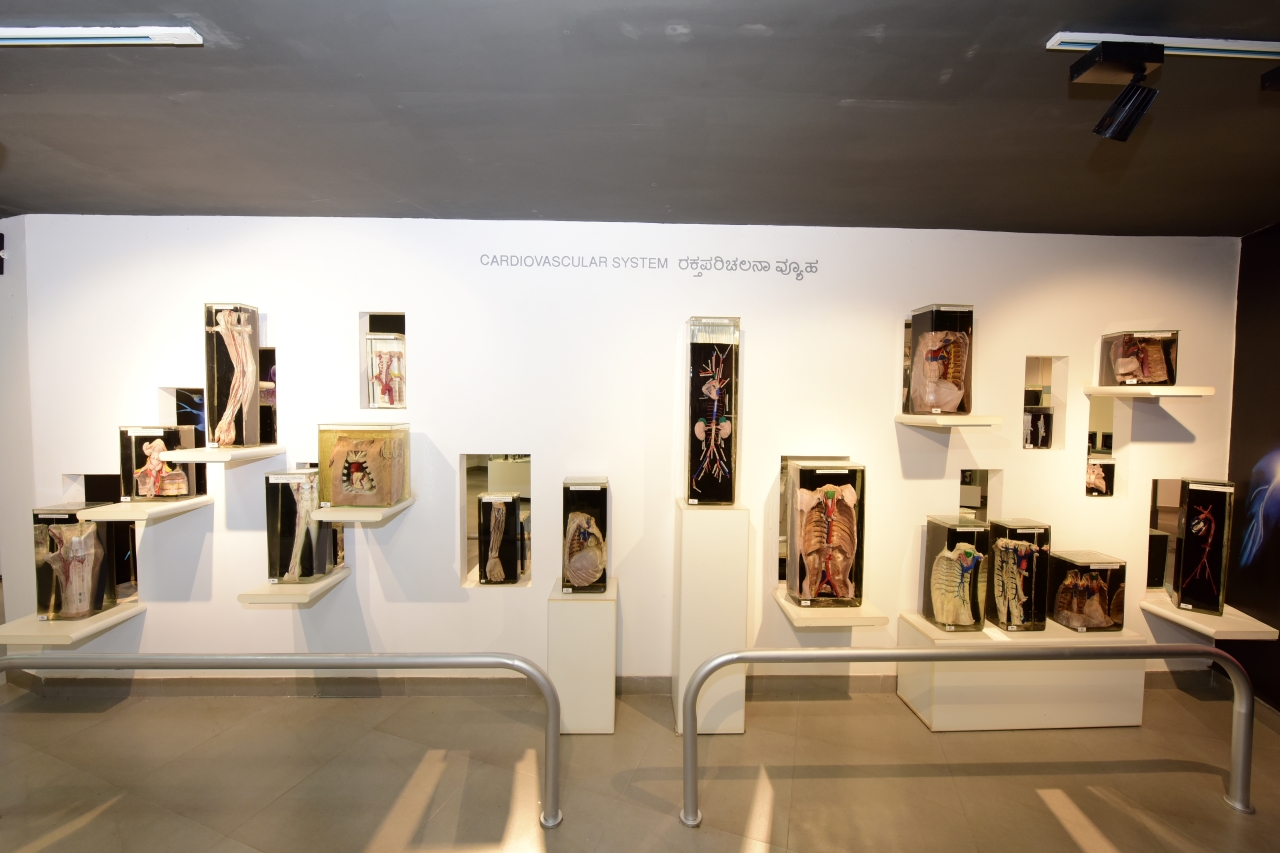
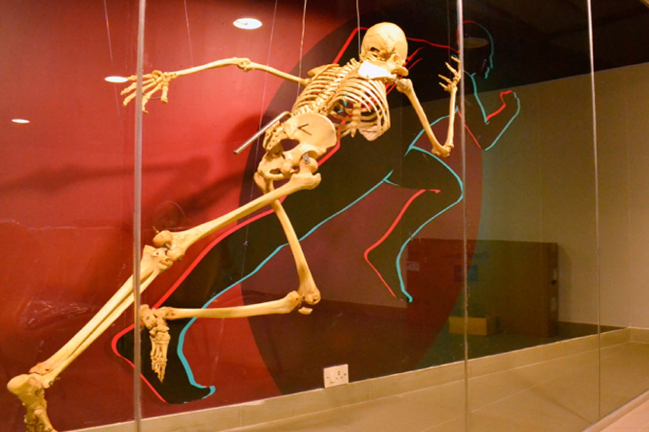
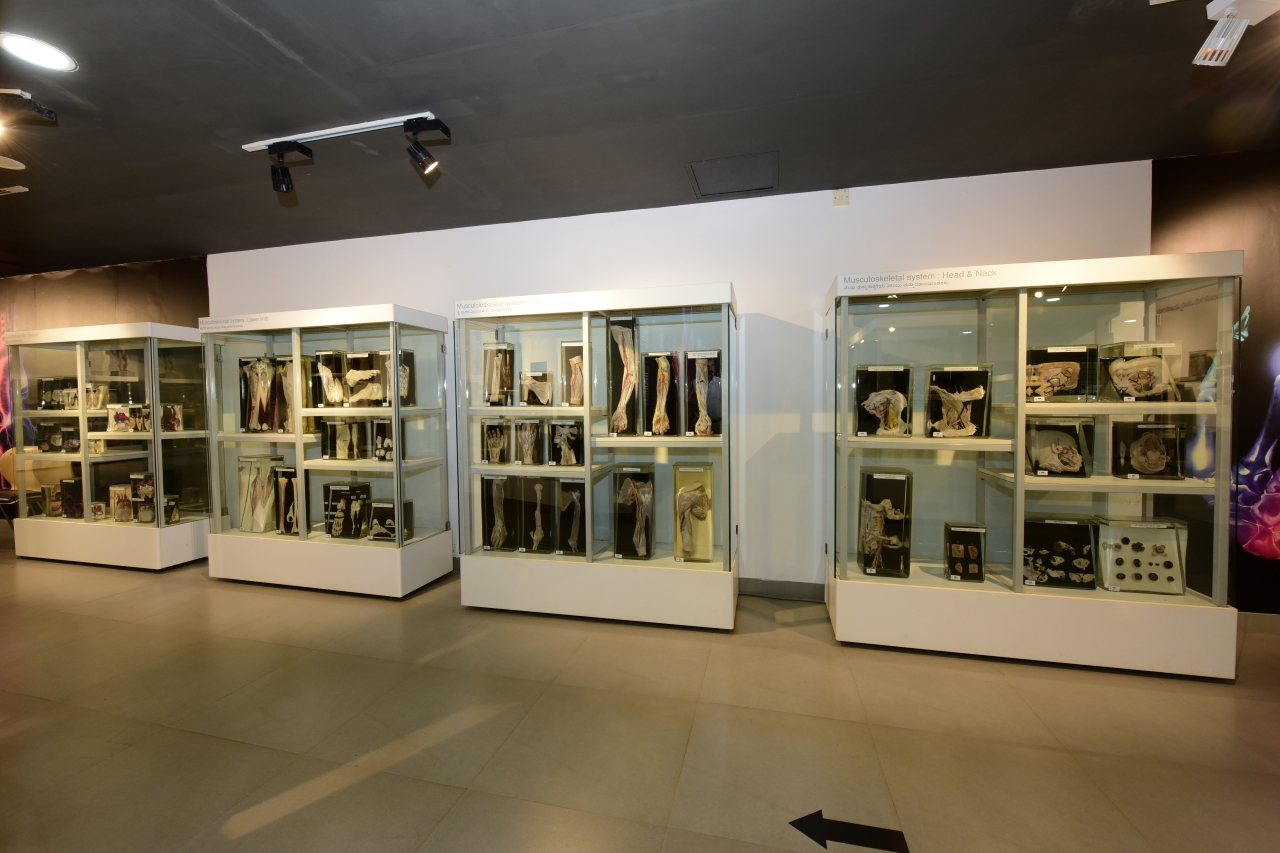
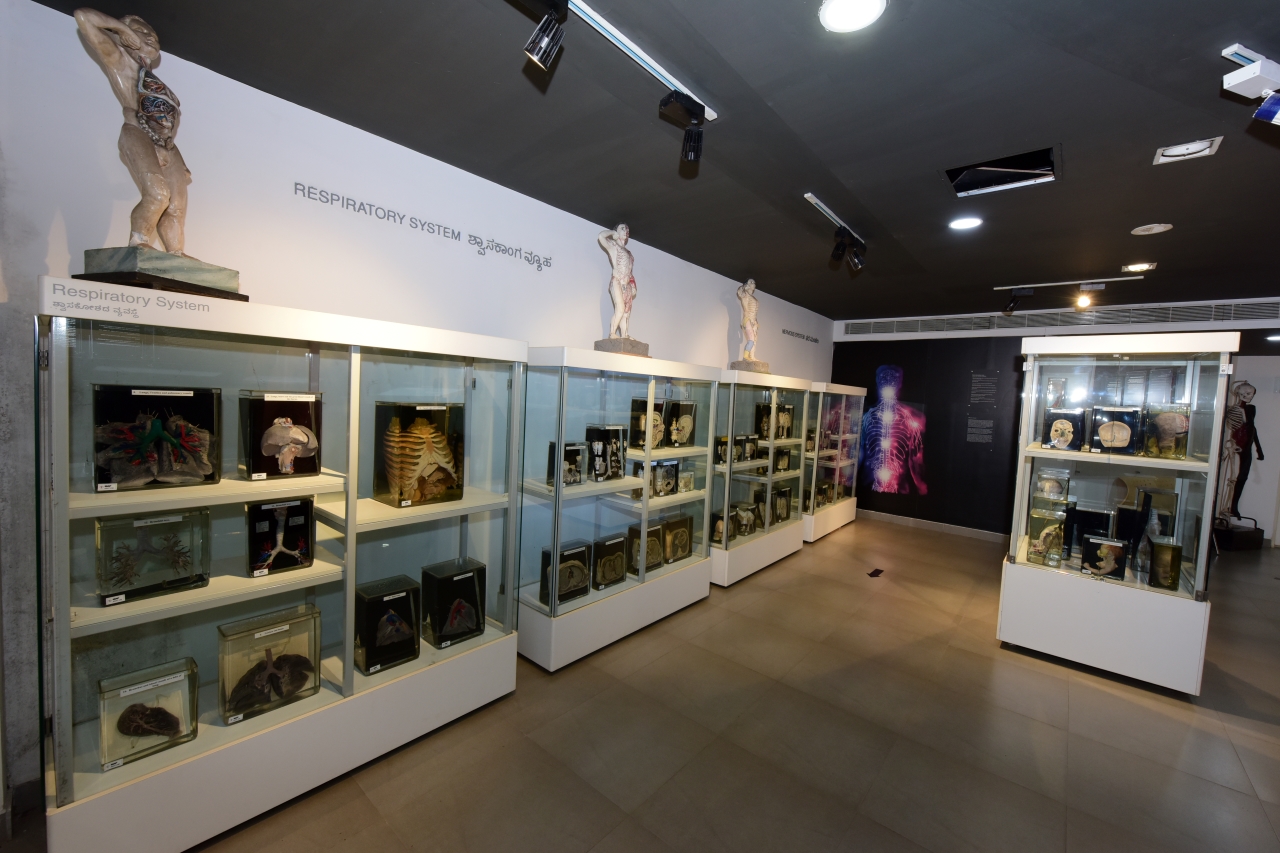
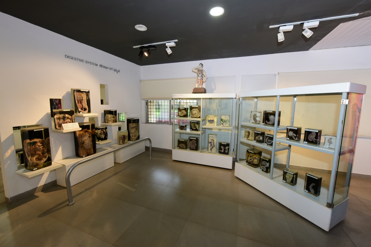
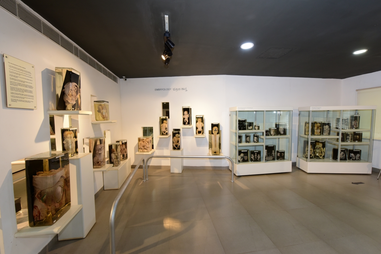
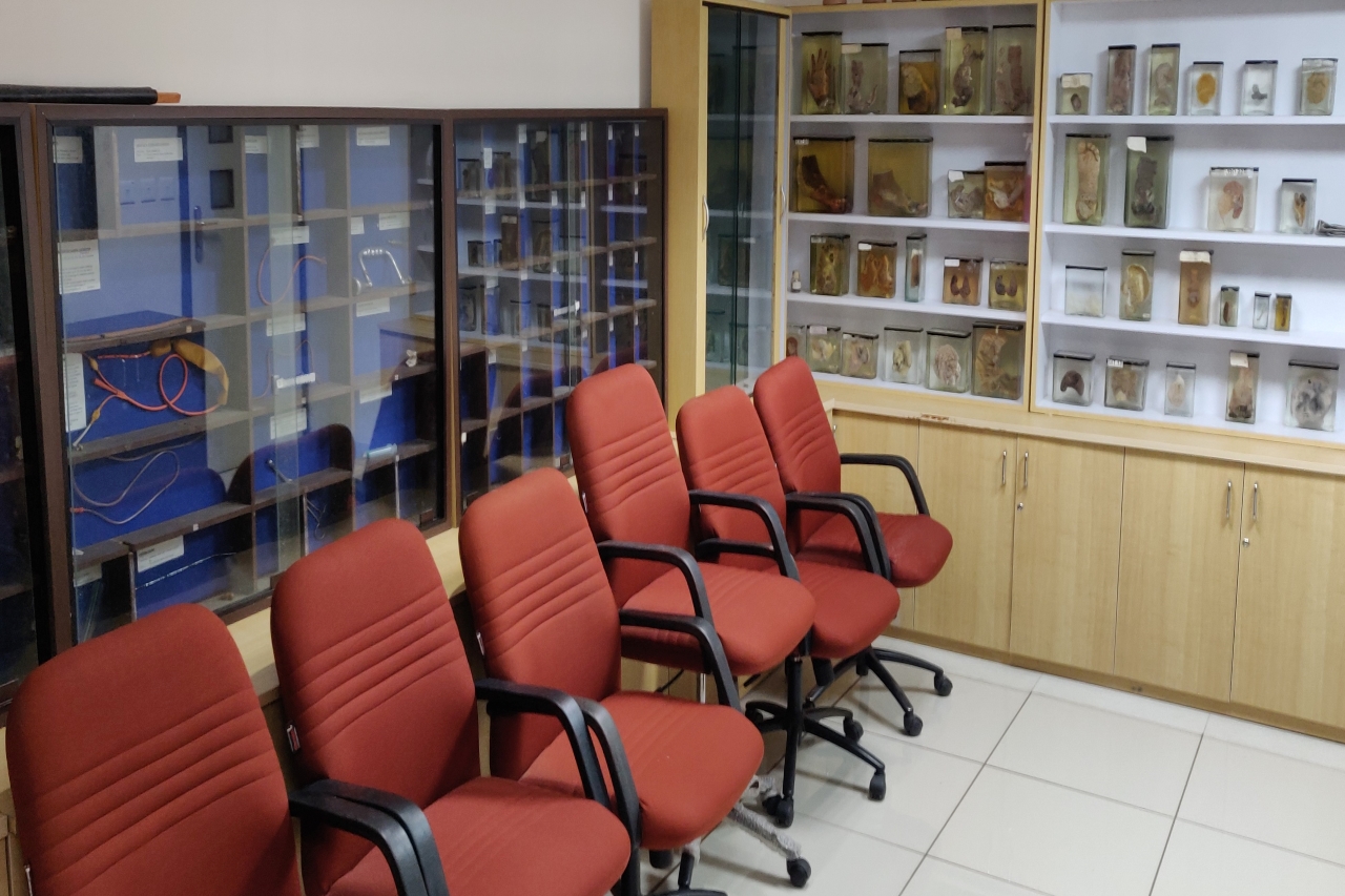
Revamped and renamed as the Manipal Museum of Anatomy and Pathology in November 2012, the sprawling Anatomy section houses well-preserved specimens of the human body from head to toe, and everything in between. A section on comparative anatomy houses the skeletons of various other animals, and carefully crafted models and charts augment the entire experience.
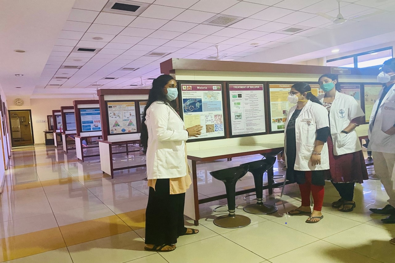
The Pathology Museum, which displays diseased body parts and organs, has a popular section on life-style related diseases, and their impact on the human body. Work to convert it into a digital or virtual museum is completed and is available on the intranet.
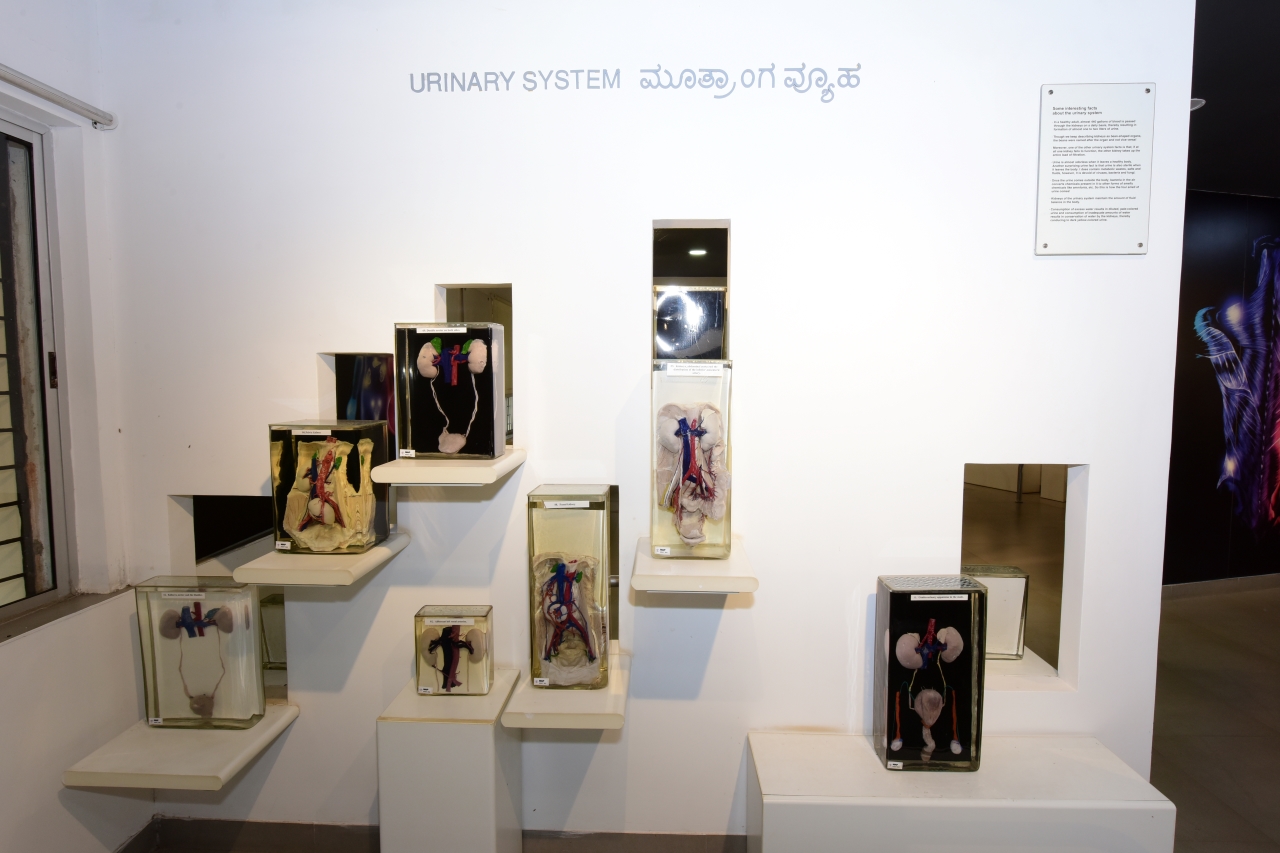
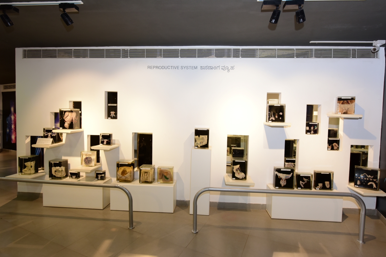
The museum is divided into two main sections: Anatomy and Pathology. The anatomy section displays specimens of normal human body parts and organs in a manner that makes them easy to understand. Each organ system of the human body can be explored as a separate entity. Every bit of the body is displayed from head to toe from different sections. There is a section on comparative anatomy, where a large collection of animals, their skeleton, and bones are displayed. The museum of Pathology displays diseased body parts and organs. The section on life-style related diseases displays specimens of diseases that occur in humans.
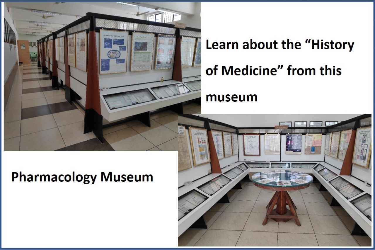
Museum of Pharmacology
The museum has a display of specimens, charts, models, and a section on "History of medicine" in addition to a computer and seating area for students.
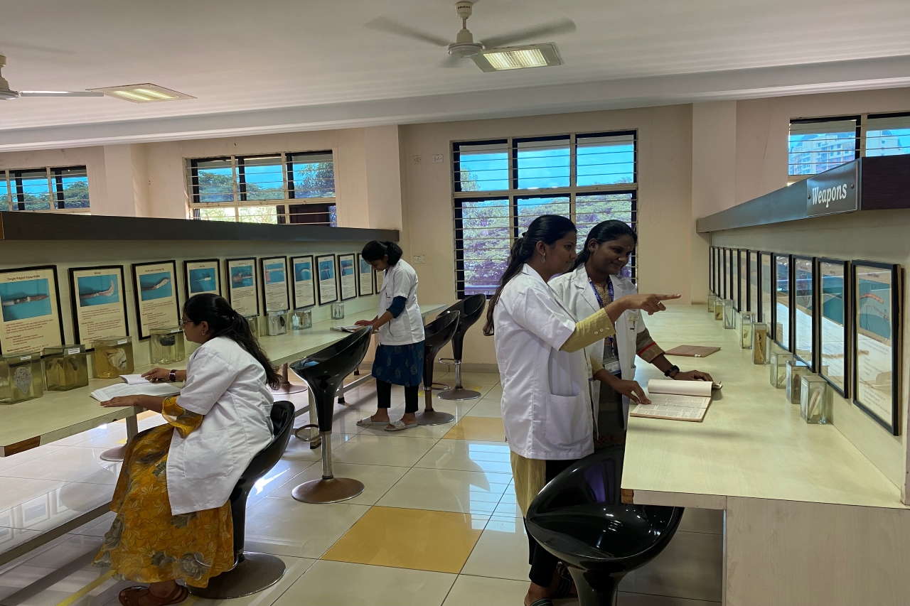
Museum of Forensic Medicine
Department museum is set up over an area of 225 sqm2 which contains mounted specimens in glass jars depicting various conditions of forensic importance. It also contains flow charts, photographs, and wax models of various aspects of forensic medicine. Prototypes of firearms and toxicological specimens are also part of the department museum. Important cases which were milestones in the history of Forensic medicine is also depicted with photographs and description. All the specimens are arranged in serial rows with supporting catalogs which give details of the specimen. There are arrangements to seat 80 students at a time.
Community Medicine Museum
The department has a museum of 175 sq. mt. with an extensive collection of 54 exhibits & 128 Charts. These exhibits involve the evolution of community medicine, concepts of health, epidemiology, communicable & noncommunicable diseases, maternal & child health, and health legislation. There are specimens of various common diseases in the local community and slides related to those. There are Photographs of eminent scientists of public health importance used for undergraduate and postgraduate teaching.
General Surgery Museum
The surgery museum houses numerous historical surgical specimens ranging from common to rare surgical diseases as well as historical devices and instruments with detailed descriptions, catering to young surgeons, keeping in mind that surgical training begins with an appreciation of its history. Integrating these specimens in teaching programs aids in nurturing young minds by providing a holistic learning environment blending history with current surgical progress. We believe that is paramount that both medical students and surgical trainees have access to these facilities to inspire and aid their academic progress.
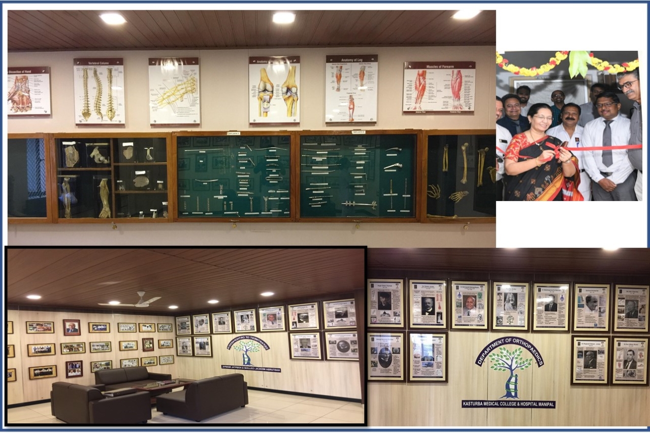
Orthopaedic Museum
The department of Orthopaedics boasts of having a museum. The museum, established in 2015, showcases a photographic depiction of the history and evolution of the department, along with archives of scientific papers and research articles published in various national and international journals by our esteemed faculty. The museum walls are decorated with posters describing the works of some of the finest Orthopaedic surgeons across the globe.