
About the Department
The Department of Radiodiagnosis & Imaging in KMC was established in 1962.Radiology as a specialty is an integral part of Imaging, diagnosis, treatment & post treatment follow up of every patient. We thrive to reduce hospital stay & accelerate recovery process.
Our department is also involved in numerous Research activities, academic pursuits, Inter-departmental teachings & collaborations. On a daily basis, 200-250 Ultrasound scans, 600 Radiographs, 140 CT scans & 60 MRI scans are performed along with Mammograms, special Radiographic procedures, Angiograms & Image guided Interventions.
Over the time, we have nurtured Musculoskeletal Imaging as a subspecialty along with Image guided Biliary, Vascular Interventions. We aim to develop these subspecialties into fellowship courses in due time.
Adjunct Faculty
- Dr. Sikandar Mohammed Shaikh is a distinguished radiologist with over two decades of clinical expertise and academic involvement in diagnostic imaging and PET-CT. Holding credentials such as DNB, MNAMS, FICR, and the prestigious EDIR certification from the European Board of Radiology.
- Dr. Sanjeev Shakhapur is an internationally trained radiologist with extensive experience in musculoskeletal imaging and sports medicine. He holds prestigious qualifications from the UK and Europe, including the FRCR, CCST, and diplomas in MSK Radiology and Sports Medicine.
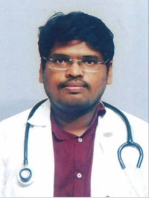
- Dr. Chandan Kakkar is a highly skilled radiologist with specialized expertise in Body Imaging. With a clinical fellowship from McGill University Hospitals, Montreal, Canada, he has gained advanced knowledge and experience in oncologic, transplant, complex hepatobiliary, and gynecological oncology imaging. H
- Dr. Anagha R. Joshi is a highly experienced radiologist with comprehensive expertise in various imaging techniques, including advanced CT imaging. As the Professor and Head of the Department of Radiology at LTMG Hospital & LTM Medical College.
- Dr. Avinash Kambadakone-Ramesh is an expert in abdominal imaging with a distinguished academic and clinical career. He completed his MD at Kasturba Medical College, Manipal, India, and is currently a faculty member in the Division of Abdominal Imaging at Massachusetts General Hospital in Boston, USA.
- Dr. Roopa Seshadri is a highly skilled radiologist with a specialized focus on neuroimaging. She completed her MD and DM from the National Institute of Mental Health and Neurosciences (NIMHANS), one of the leading institutions in the field of neuroscience in India.
Alumni
Name |
Year of passing |
Current position |
Dr Naveen M Mulimani |
2009 |
Associate Professor, Jawaharlal Nehru, Belgaum.
|
Dr Ullas V A |
|
Consultant – Neuroradiology, Manipal Hospitals, Bangalore |
Dr. Lakshmikanth H K |
2013 |
Associate Professor, Kanachur Institute of Medical Sciences, Mangalore |
Dr Samir P M |
2014 |
Consultant Musculoskeletal Radiologist, York and Scarborough Teaching Hospitals, UK |
PhD Awarded/Ongoing
S. no |
Candidate name |
Year |
1. |
Dr Suresh Sugumar |
2017 |
2. |
DR Rahul P Kotian |
2020 |
3. |
Abhimanyu Pradhan |
2018 |
4. |
Shree Kripa |
2019 |
5. |
Nitika Panakkal |
2019 |
6. |
P Vaidehi Nayantara |
2019 |
7. |
Priyanka Shridharan |
2019 |
Services Offered
The department is well-equipped with state-of-the-art machines, manned by competent radiologists and well-trained technologists. It has been a pioneer in diagnostic and interventional radiology and has several laurels to its credit. It was the first to get a CT scan machine in Karnataka and second institute in South India to have a PACS (Picture archiving and communication system). The department is suitably equipped to provide almost all possible diagnostic and therapeutic radiology services for both inpatients and outpatients.
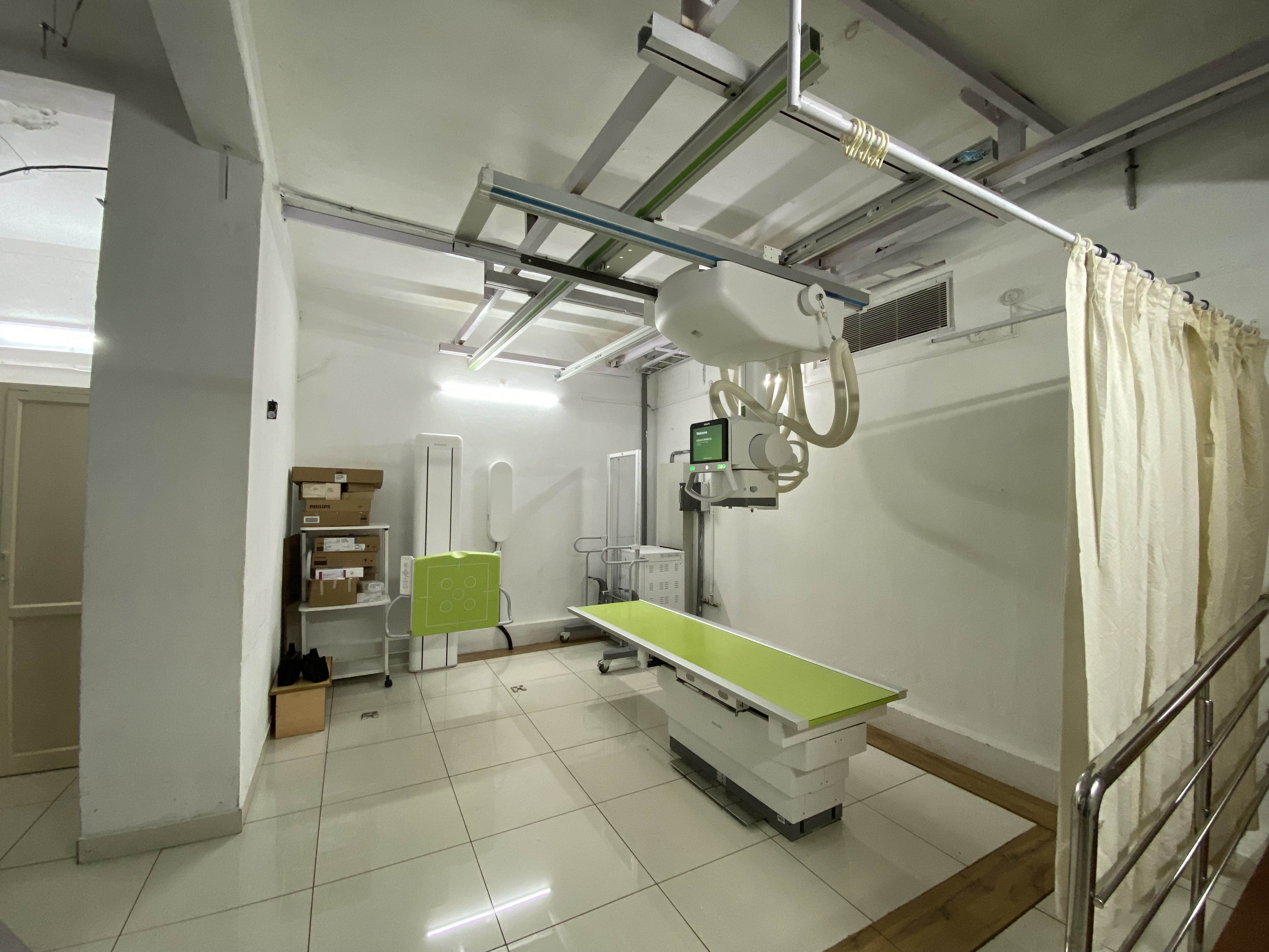
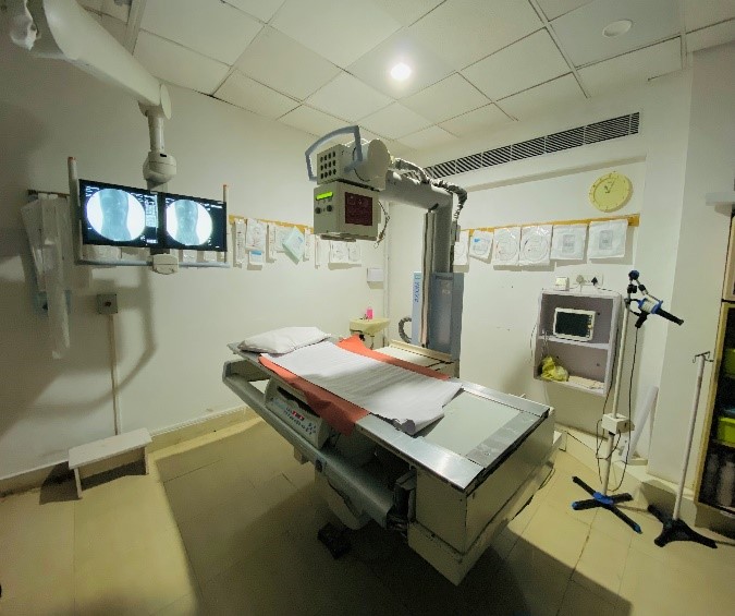
RADIOGRAPHY
For routine radiography of the chest, abdomen and skeletal system, both Computed Radiography (CR) and Digital Radiography (DR) are utilized.
Ten portable units are available 24 x 7 for emergency scanning in the ICU and for trauma cases.
Several fluoroscopy-guided diagnostic and interventional procedures using contrast media are performed on the Siemens Iconos R200 machine, including Barium studies, hysterosalpingograms, IVU and MCU, sialograms, T-tube cholangiograms, percutaneous transhepatic biliary drainage (PTBD) and percutaneous nephrostomy (PCN).
All routine radiographs and contrast procedures are acquired in digital format, uploaded to PACS (Picture Archival and Communication System) and reported with the help of sophisticated Barcos.
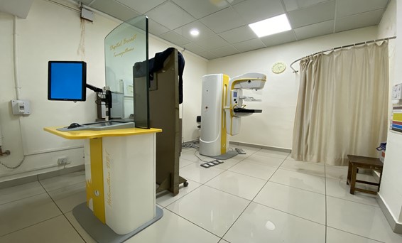
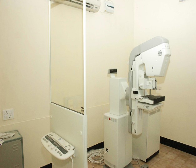
DIGITAL MAMMOGRAPHY
Mammography screening entails examining thousands of healthy women. Therefore, it is of utmost importance to keep the radiation dose as low as possible. However, high image quality is indispensable to be able to detect small lesions. The modern Metaltronica Helianthus DBT machine enables the radiologist to take images of superior quality without compromising on radiation safety. The detection and characterization of smaller lesions is further augmented by utilizing sonography and giving a combined sono-mammography report.
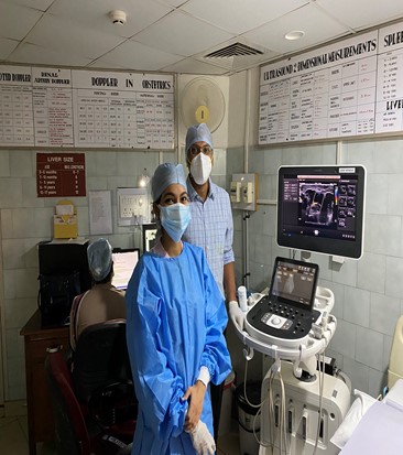
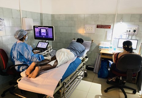
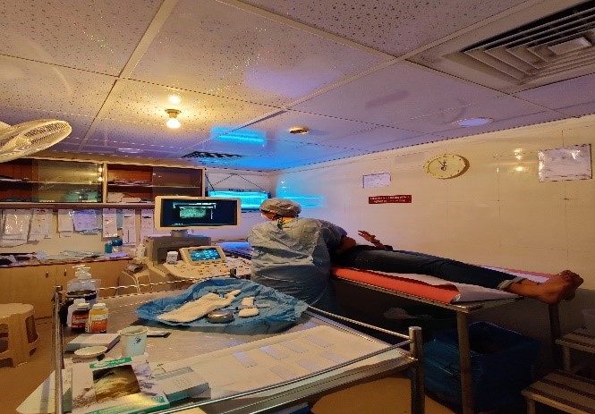
ULTRASONOGRAPHY AND COLOUR DOPPLER
Eleven sophisticated ultrasound machines are used for both diagnostic and therapeutic procedures.
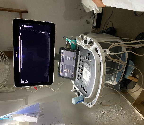
Seven machines are used for examination of the abdomen, pelvis and small parts, and for Colour Doppler to evaluate patients for deep venous thrombosis, varicose veins and peripheral vascular disease.
One state-of-the-art system is utilized for performing ultrasound-guided procedures, such as FNAC, biopsy and fluid aspiration.
The three portable units are used primarily for emergency scanning of ICU patients and bedside image-guided procedures, like pigtail catheter insertion and fluid / abscess aspiration. They are also utilized for intra-operative assistance to colleagues in other departments, such as for insertion of peripheral lines and reduction of zygomatic fractures.
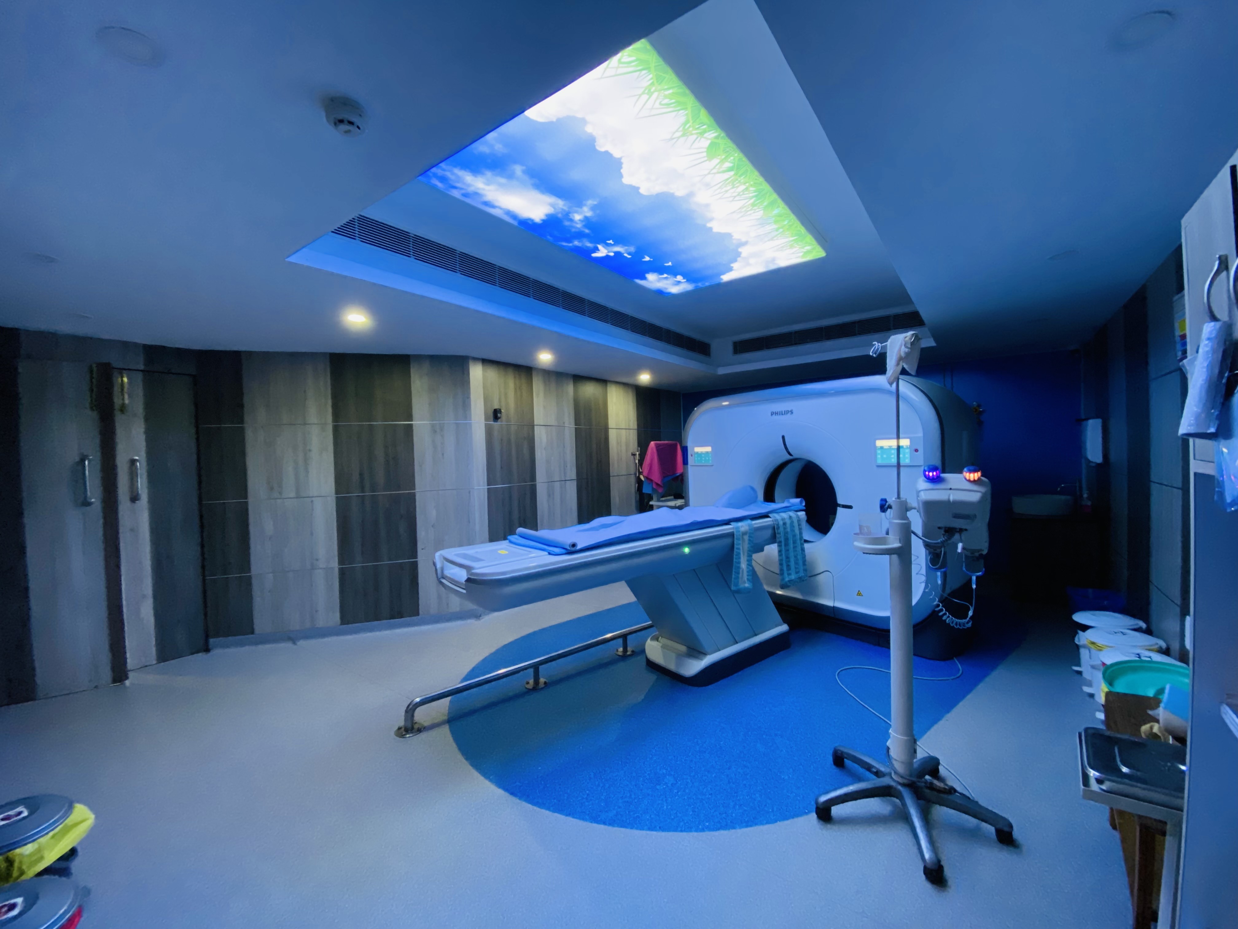
COMPUTED TOMOGRAPHY
State-of-the-art 128-slice Philips Incisive CT scanner and 16-slice Philips Brilliance Big-Bore scanner with 3D rendering capabilities are utilized for both routine and emergency scans, done 24 x 7.
Angiography scans with 3D reconstruction performed, including cerebral, peripheral limb, abdominal, aortogram and pulmonary. Special features include coronary angiography and CT guided procedures, including biopsies, radiofrequency tumour ablation, nerve blocks and sclerotherapies.
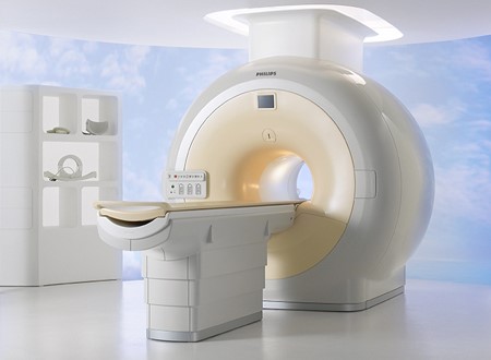
MAGNETIC RESONANCE IMAGING
United imaging 3T and Philips ACHIEVA 1.5T machines are utilized for routine imaging of the brain, spine, abdomen and pelvis, and musculoskeletal system, including lumbar and brachial plexus studies.
Advanced techniques include proton spectroscopy for evaluation of brain tumours, cardiac MRI, breast MRI and fetal MRI.
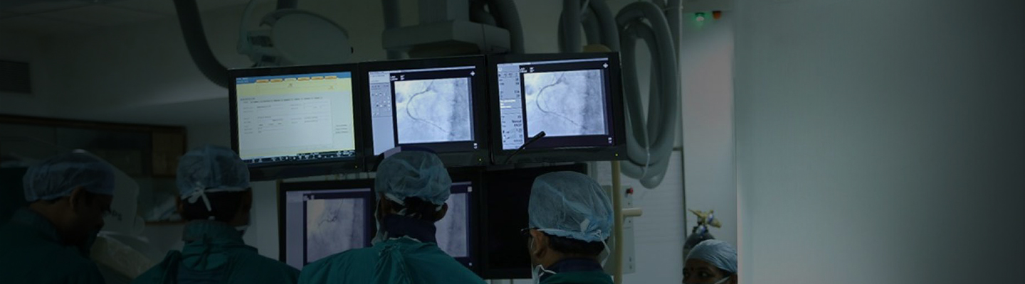
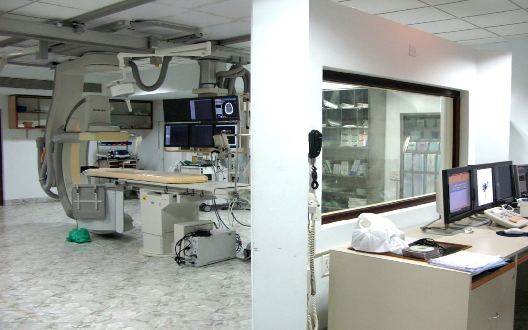
INTERVENTIONAL RADIOLOGY
Minimally invasive and targeted therapies are used to treat patients and help their early recovery. The instruments for the procedures are utilized under imaging guidance:
Fluoroscopy guided diagnostic procedures:
Barium studies
Hysterosalpingography
IVU, MCU
Fistulogram
T-Tube cholangiogram
Digital Subtraction Angiography (DSA)
Fluoroscopy guided therapeutic procedures:
Percutaneous transhepatic biliary drainage (PTBD)
Percutaneous nephrostomy (PCN)
Sclerotherapy
Trans arterial chemoembolization (TACE)
Uterine artery embolization
Ultrasound guided procedures:
FNAC
Biopsy
Pleural tap
Ascitic tap
Aspiration of abscess
Pigtail catheter insertion
CT guided procedures:
FNAC / Biopsy
Pigtail catheter insertion
Radiofrequency ablation
Nerve blocks
EMERGENCY SERVICES
Round-the-clock services are provided on emergency basis. Angiography and embolization for bleeding Catheter drainage for abscesses and obstructive uropathy
Research
Research is one of the main aspects of routine department work in addition to the clinical activities. Post graduates carry out thesis research work under the guidance of professors. For this, state-of-the-art imaging equipments are available, like MRI, MDCT, digital mammography and hi-end ultrasound equipment.
Several PhD scholars (10 students) are also enrolled with various research projects in the department. One of them is a full-time PhD student and the others are part-time.
Department also possess Medical Imaging Research suite (MIRS) where the students with a technical background from MIT carry out the research work. Few PhD students are also stationed here. Some of the objectives of the MIRS include:
1. Ffacilitate students & faculty to carry research in the area of medical imaging & bring out publications, patents & products.
2. To train professionals, faculty in the area of clinical domain with industry support.
3. To collaborate with academic sites working in similar area.
4. To offer full-time student internships/projects, thereby providing an opportunity to work on Health Care projects under the guidance of Radiologists & engineers. This prepares them better for jobs in the healthcare market.
Ongoing research:
Sl No |
Title of the grant |
Granting agency |
Principal Investigator |
Duration of the project |
|
1 |
Modelling an Accurate for Detection and Quantitative Assessment of Malignant Tumors in pre and post therapy Spinal Metastases using PETCT |
DST –SYST scheme |
Dr Prakashini K – co -PI |
|
|
2 |
• Mid-cycle MRI to predict Complete Pathologic Response to Neoadjuvant Chemotherapy in Breast Cancer patients |
Dr Smiti Sripathi Endowment fund |
Dr Aanthriksha K |
2020-2023 |
· Role of DTI in Parkinsonism disorders
· Predicting Histopathological and Molecular Subtype of Non- Small Cell Lung Carcinoma based on CT features
· Diffusion Tensor Imaging - MR Tractography in Stroke Patients to Predict Motor Outcome
· Subtraction imaging in nonvascular MRI.
· Role of MRI in the assessment of patients with anterior Knee pain with emphasis on Patellar instability and impingement syndromes
· Association between muscle strength and ultrasound measured muscle thickness and echo intensity in South Indian Individuals with and without hypothyroidism
· Spectrum of CT findings in patients of COVID-19 and it’s complications
· Multiparametric MRI for Bladder Cancer Using VI-RADS for the Detection of Detrusor Muscle Invasion: An Observational Study to check the Reliability of VI-RADS scoring in Indian Population
· Utility of multiphase CT cerebral angiography in acute ischemic stroke
· Mid-cycle MRI to predict Complete Pathologic Response to Neoadjuvant Chemotherapy in Breast Cancer patients
· Role of Shear wave elastography in assessing tendinopathy of supraspinatus tendon and muscle tendoachiliies and common extensor tendon of elbow in an Indian population.
· Evaluating the accuracy of ABC/2 volume formula compared to computer assisted volumetric analysis of intracranial haemorrhages on computed tomography.
· MRI based measurements of tibial and femoral parameters on an Indian population with insufficient knees.
· Role of MRI in staging of rectal cancer and value of gadolinium enhanced MRI in predicting the extent of rectal cancer.
· Detection and Staging of Laryngeal cancer Using Machine Learning and Radiomics.
· Optimisation of b-value in diffusion weighted MRI for benign and malignant breast lesions
· Development of an analytics algorithm to predict clinical outcomes of Head and Neck Cancer using features and randomics.
· Development and testing of dose reduction strategies on radiation dose and image quality in CT angiography
· Establishing diagnostic reference levels and optimisation of radiation dose for computed tomography of head in paediatric population
· Variations in Default Mode Network of Brain Among Raja Yoga Meditators, Non-Meditators and Patients with Migraine Disorder – An in Depth Vexel Based Morphometric Analytical Study of sMRI and fMRI
· Establishing diagnostic reference levels and optimization of radiation dose for head and neck and pelvic computed tomography protocols in radiation therapy planning
· Influence of low kilo voltage protocol on image quality, iodine and radiation dose for abdominopelvic computed tomography.
· Computer aided Diagnosis system for detection and classification of liver tumor using Computed Tomography Images
· Prognostication of Acute Ischemic Stroke (AIS) in anterior cerebral circulation, based on the distribution of cerebral collaterals and clot burden on CT Angiography studies.
· Prognostication of Acute ischemic stroke by developing a prognostic index based on a clinical characteristics and stroke volume
· Artificial intelligence enabled acute ischemic stroke infarct volume estimation and intracranial emergent large vessel occlusion quantification
· Development and testing of optimal cardiac CT protocol using fixed time delay for congenital heart disease in children.
· Modelling of textures-based assessment system to accurately determine response to therapy of vertebral metastasis on DWI IN COMPETENCE WITH PET CT
· Impact of yoga on central and Peripheral vascular function and cognitive functions among the desk-based workers.
· Exploring application of radiomics in brain tumour assessment and treatment.
Publications
1. Divya B. a,∗ , Rajesh Parameshwaran Nair b , Prakashini K. c , Girish Menon R. b , Paul Litvak d , Pitchaiah Mandava d , Deepu Vijayasenan e , Sumam David S. e; Generalizable DNN model for brain tumor sub-structure segmentation from low-resolution 2D multimodal MR Images; Biomedical Signal Processing and Control 100 (2025) 106916; https://doi.org/10.1016/j.bspc.2024.106916 -Q1
2. Sanjeev Kumar1 · K. Devaraja2 · Jasbon Andrade3,7 · Deepak Nayak4 · Geetha Vasudevan4 · Suresh Pillai2, K. Prakashini3 · P. S. Priya3 · Kinjal Shankar Majumdar2 · Naveena AN Kumar5 · Nawaz Usman5 · Adarsh Kudwa6, Kailesh Pujary1 · Balakrishnan Ramaswamy1 · Dipak Ranjan Nayak1,8 Computed Tomography in Predicting the Mandibular Involvement by Oral Cancer, a Retrospective Study ; Indian Journal of Otolaryngology and Head & Neck Surgery (2025) 77:1281–1288; 10 January 2025-https://doi.org/10.1007/s12070-025-05321-x - Q3
3. Praveen Kumar Tirlangi, Swathi Kiran, Vandana KE, C Mukhopadhyay, Ramit Kundu, Ananth Pai, Priya PS, Kavitha Saravu; Adjunctive nivolumab in combination with antibiotic therapy for the management of refractory melioidosis in a patient with metastatic breast cancer and chemotherapy-induced pancytopenia; Transactions of The Royal Society of Tropical Medicine and Hygiene, 2025; trae142, https://doi.org/10.1093/trstmh/trae142 -Q2
4. Divya Josephraj1 , Ravindranath Vineetha1 , Priya Pattath Sankaran2 , Prakashini Koteshwara2 , Mathangi Kumar1 , Kalyana Chakravarthy Pentapati3; Impact of Artifacts Caused by Intraoral Dental Materials in Magnetic Resonance Imaging; Pesquisa Brasileira em Odontopediatria e Clínica Integrada 2025; 25:e240044; Q2- https://doi.org/10.1590/pboci.2025.057
5. Poorthy Torne1 , Dasharathraj k. Shetty2*, Krishnamoorthi Makkithaya3 , Prasiddh Hegde4 , Manu Sudhi5 , Phani Kumar Pullela1 , Tamil Eniyan t6 , Ritesh Kamath2 , Staissy Salu7 , Pranav Bhat1 , Girisha s2 , Priya P S 8; VGG-16, VGG-16 With Random Forest, Resnet50 with SVM, EfficientNetB0 with XGBoost-Enhancing Bone Fracture Classification in X-Ray Using Deep Learning Models IEEE Access; DOI 10.1109/ACCESS.2025.3534818 -Q1
6. Nagendra D, Chaudhuri S, Gupta N, Shanbhag V,Eshwara VK, Rao S, Priya P S,et al. Prevalence, Risk Factors, and Clinical Outcomes of Hypervirulent Klebsiella pneumoniae Strains among Klebsiella pneumoniae Infections: A Systematic Review and Meta-analysis. Indian J Crit Care Med 2025;29(4):370–393- Q2
7. Tulasiram Bommasamudram, Zoe G. Morrell, Matthew J. Clarkson, Kirtana Raghurama Nayak, Rajagopal Kadavigere, Aaron P. Russell & Stuart A. Warmington (05 Mar 2025): Chronic adaptations to blood flow restriction aerobic or bodyweight resistance training: A systematic review, Journal of Sports Sciences, DOI: 10.1080/02640414.2025.2474346 https://doi.org/10.1080/02640414.2025.2474346 -Q1
8. Tulasiram Bommasamudram, Kirtana Raghurama Nayak, Matthew J. Clarkson, Rajagopal Kadavigere, Aaron P. Russell & Stuart A. Warmington (03 Mar 2025): Acute responses to aerobic and bodyweight exercises with and without blood flow restriction in sedentary individuals – A randomized crossover study, Journal of Sports Sciences, DOI: 10.1080/02640414.2025.2474356; https://doi.org/10.1080/02640414.2025.2474356 -Q1
9. Shreya, Lydia Shobha Andrade, Rajagopal K V, Vikram Palimar, Muhammad Nasir Ahmad, Vinod C. Nayak, Varun Kumar S G, Bhukya Nom Kumar Naik; Cervical vertebral metrics’ A reliable approach to forensic identification: A comprehensive review2025, Translational Research in Anatomy, p. 100391; DOI;10.1016/j.tria.2025.100391 -Q2
10. KRISHNAPRASAD PERUVAJE RAMAKRISHNA, Ruchitha, Rajgopal, hitesh, Monappa, Lakshmiha, Chinmaya: The Radiological evaluation and comparison, between standard and hindfoot alignment parameters in Adult Acquired Flatfoot Deformity in Indian population : Radiological assessment of hind foot alignment in adult acquired flat foot;
11. P Vaidehi Nayantara1· Surekha Kamath1 · Manjunath KN2 · Rajagopal Kadavigere3; Computer‑Aided Diagnosis System for Classifying the Liver Lesions from Multiphase CT Images ; Sensing and Imaging; Volume 26, Issue 1 ; https://doi.org/10.1007/s11220-025-00568-8
12. Dkhar W, Kadavigere R, Ravichandran S et al. Hormonal Influences on ADC Values in Breast Tissues: A Scoping Review of DWI in Pre- and Post-menopausal Women [version 3; peer review: 2 approved]. F1000Research 2025, 13:857 (https://doi.org/10.12688/f1000research.153999.3)
13. Mohamed Sajer R, Pendem S, Kadavigere R et al. Applications of MR Finger printing derived T1 and T2 values in Adult brain: A Systematic review [version 1; peer review: 2 approved]. F1000Research 2025, 14:54 (https://doi.org/10.12688/f1000research.160088.1)
14. PoovithaShruthi Paramashiva a 1, Annapoorna K a 2, Vaishali K b 3, Baskaran Chandrasekaran c 4, Shivashankar K.N. d, Suresh Sukumar e 5, Sneha Ravichandran e 6, Dilip Shettigar e 7, Sathya Sabina Muthu e 8, Koustubh Kamath e 9, Rajagopal Kadavigere f 10 ; Enhancing well-being at work: Qualitative insights into challenges and benefits of long-term yoga programs for desk-based workers; Advances in Integrative Medicine, https://doi.org/10.1016/j.aimed.2025.01.001
15. Abhimanyu Pradhan, M.Sc.1 , Rajagopal Kadavigere, M.D.2 , Suresh Sukumar, Ph.D.1; Establishing Local Diagnostic Reference Levels for CT Angiography Examinations; Journal of Health Science & Medical Research; DOI 10.31584/jhsmr.20241084; Volume 43, Issue 1Jan-Feb 2025
16. Barnes NA, Dkhar W, Kadavigere R et al. Exploring mean kurtosis in MR diffusion kurtosis imaging for early detection of lumbar spine degeneration: a systematic review [version 1; peer review: awaiting peer review] F1000Research 2025, 14:440 https://doi.org/10.12688/f1000research.163638.1;
17. Nicolet, Mariaa;Priyanka a;Kadavigere, Rajagopal b;S Nayak, Shailesha;Pendem, Saikirana.;Aggarwal, Surbhi Guptab;Pires, Tanciaa;Varsha R.a; Comparison of sagittal measurements of cervical spine in spondylosis patients between Magnetic Resonance Imaging and Radiograph; F1000Research; (https://doi.org/10.12688/f1000research.159504.1)https://eprints.manipal.edu/view/subjects/RAD.html
2024:
1. Shruthi P P, Kamath K, K V, KN S, Sugumar S, Ravichandran S, David LR, Hogg P, V G, Kadavigere, R* B, Saha S. Impact of yoga on the central and peripheral vascular function among desk-based workers: A single-centred trial study. F1000Research. 2024 Apr 15;13:277. -Q1 https://doi.org/10.12688/f1000research.135239.2
2. Priyanka, Kadavigere R, Sukumar S. Low Dose Pediatric CT Head Protocol using Iterative Reconstruction Techniques: A Comparison with Standard Dose Protocol. Clinical Neuroradiology. 2024 Mar;34(1):229-39. -Q1 https://doi.org/10.1007/s00062-023-01361-4
3. Obhuli Chandran M, t1, Saikiran Pendem 1, Priya P S, 2, Cijo Chacko, Priyanka, Rajagopal Kadavigere* Influence of deep learning image reconstruction algorithm for reducing radiation dose and image noise compared to iterative reconstruction and filtered back projection for head and chest computed tomography examinations: a systematic review [version 1; peer review: awaiting peer review]. F1000Research 2024, 13:274; Q1 https://doi.org/10.12688/f1000research.147345.1
4. Chandran M O, Pendem S, Priya P S 2, Cijo Chacko 3, Priyanka 1, Rajagopal Kadavigere 2. Comparison of image quality between Deep learning image reconstruction and Iterative reconstruction technique for CT Brain- a pilot study [version 1; peer review: 4 approved]. F1000Research 2024, 13:691 -Q1 (https://doi.org/10.12688/f1000research.150773.1)
5. K B, S., Vaishali, K., Kadavigere, R. et al. Effects of high-intensity interval training versus moderate-intensity continuous training on vascular function among individuals with overweight and obesity—a systematic review. Int J Obes (2024).Q1 https://doi.org/10.1038/s41366-024-01586-4
6. Dhamija A, Andrade LS, Prakashini K and Gupta C*. Correlation of age with the size of subcortical nuclei of the brain and its implication in degenerative disease: A magnetic resonance imaging study [version 2; peer review: 2 approved]. F1000Research 2024, 12:1230- Q1 https://doi.org/10.12688/f1000research.139515.2
7. Gorthi SP, Suresh K, Koteshwara P. Association of Internal Cerebral Vein Asymmetry and Collateral Status with Outcome of Anterior Circulation Acute Ischemic Stroke Cases (P3-5.028). In Neurology 2024 Apr 14 (Vol. 102, No. 17_supplement_1, p. 3927). -Q1; https://doi.org/10.1212/WNL.0000000000205466
8. Dkhar W, Kadavigere R, Ravichandran S et al. Hormonal Influences on ADC Values in Breast Tissues: A Scoping Review of DWI in Pre- and Post-menopausal Women [version 2; peer review: 1 approved]. F1000Research 2024, 13:857 – Q1 https://doi.org/10.12688/f1000research.153999.2
9. Divya B. a,∗ , Rajesh Parameshwaran Nair b , Prakashini K. c , Girish Menon R. b , Paul Litvak d , Pitchaiah Mandava d , Deepu Vijayasenan e , Sumam David S. e; Generalizable DNN model for brain tumor sub-structure segmentation from low-resolution 2D multimodal MR Images; Biomedical Signal Processing and Control 100 (2025) 106916-Q1; https://doi.org/10.1016/j.bspc.2024.106916
10. Priyanka, Kadavigere, R., Nayak S, S., Chandran M, O., Shirlal, A., Pires, T., & Pendem, S. (2024). Impact of artificial intelligence assisted compressed sensing technique on scan time and image quality in musculoskeletal MRI – A systematic review. Radiography, 30(6), 1704-1712.Q1- DOI:10.1016/j.radi.2024.08.012
11. Kundu S, Nayak K, Kadavigere R et al. Evaluation of positioning accuracy, radiation dose and image quality: artificial intelligence based automatic versus manual positioning for CT KUB [version 1; peer review: 2 approved]. F1000Research 2024, 13:683 (https://doi.org/10.12688/f1000research.150779.1)- Q1
12. Varughese, N. A., Panakkal, N. C., Nair, V. T., Kadavigere, R., Lakshmi, V., & Sukumar, S. (2024). Effect of patient characteristics on aortic attenuation in iodinated contrast-enhanced Abdominopelvic CT: A retrospective study. Radiography, 30, 94-101. Q1 https://doi.org/10.1016/j.radi.2024.07.012
13. Sneha Guruprasad Kalthur a, Rajagopal Kadavigere b, Vrinda, Hari Ankolekar a, Dhiren Punja c, Rohini Punja a; A comprehensive morphometric analysis of superior and inferior mesenteric arteries using cadaveric dissection and MDCT angiography; Translational Research in Anatomy; Q2
http://dx.doi.org/10.1016/j.tria.2024.100328
14. Rao S, Sharan K, Chandraguthi SG, Dsouza RN, David LR, Ravichandran S, Mustapha MT, Shettigar D, Uzun B, Kadavigere R, Sukumar S, Ozsahin DU. Advanced Computational Methods for Radiation Dose Optimization in CT. Diagnostics (Basel). 2024 Apr 29;14(9):921 Q2. https://doi.org/10.3390/diagnostics14090921 .
15. Nayantara, P.V., Kamath, S., Kadavigere, R. et al. Automatic Liver Segmentation from Multiphase CT Using Modified SegNet and ASPP Module. SN COMPUT. SCI. 5, 377 (2024). – Q2 https://doi.org/10.1007/s42979-024-02719-2
16. Shruthi, Poovitha P1; Chandrasekaran, Baskaran2,3; Vaishali, K4; Shivashankar, K N5; Sukumar, Suresh6; Ravichandran, Sneha6; Kadavigere, Rajagopal7. Effect of physical activity breaks during prolonged sitting on vascular outcomes: A scoping review. Journal of Education and Health Promotion 13(1):294, August 2024. Q2 https://doi.org/10.1016/j.aimed.2024.10.011
17. Pulagam Vamsidhar Reddy & Priya PS (2024) ABC/2 formula versus computer-assisted analysis in calculating intra-cranial haemorrhage volume on computed tomographic imaging, Computer Methods in Biomechanics and Biomedical Engineering: Imaging & Visualization, 12:1, -Q2 https://doi.org/10.1080/21681163.2024.2327416
18. Nikhil Kumar1 , Pulagam Vamsidhar Reddy2 , Prakashini K2 , Priya PS 2 *, Arun Chawla3 , Ranjitha S Shetty4 , Basima Maisoon5;Size-Specific Dose Estimate – A Tool for Radiation Dose Quantification and Assessment Among Renal Colic Patients in a Tertiary Care Hospital in Southern India; International Journal of Pharmaceutical and Clinical Research 2024;16(2); 1531-1538 - Q2 – FEB - IJPCR,Vol16,Issue2,Article253.pdf
19. Praveen Kumar Tirlangi, Swathi Kiran, Vandana KE, C Mukhopadhyay, Ramit Kundu, Ananth Pai, Priya PS, Kavitha Saravu, Adjunctive nivolumab in combination with antibiotic therapy for the management of refractory melioidosis in a patient with metastatic breast cancer and chemotherapy-induced pancytopenia, Transactions of The Royal Society of Tropical Medicine and Hygiene, 2025;, trae142, Q2 https://doi.org/10.1093/trstmh/trae142
20. Shaifali1 , KV Rajagopal2 , Mithun Shekar3 , Ashok Reddy Datla4 , Pulagam Vamsidhar Reddy5 , Tanushree Kamath*6; Applications of T1 Subtraction Imaging in Non – Vascular Magnetic Resonance Imaging; Library Progress International; Vol.44 No.3, Jul-Dec 2024: P.4763-4770; Q2 https://doi.org/10.48165/bapas.2024.44.2.1
21. Abhimanyu Pradhan1 , Rajagopal Kadavigere2, *, Suresh Sukumar1 , Winniecia Dkhar1; LOW TUBE VOLTAGE CT LOWER LIMB ANGIOGRAPHY WITH AORTOGRAM: EFFECT ON RADIATION DOSE AND IMAGE QUALITY; Bradleya;Q2- https://doi.org/10.61586/1zajv
22. Laxmikant G. Keni a, Satish Shenoy B a, Chethan K , N a, Padmaraj Hegde b, Prakashini K c, Masaaki Tamagawa d, Divya D. Shetty a, Mohan Futane e, Mohammad Zuber;A Impact of obstruction size on ureter dynamics: A computational investigation; Results in Engineering; Q2 https://doi.org/10.1016/j.rineng.2024.102217
23. Nayak, S. S., Pendem, S., Menon, G. R., Sampathila, N., & Koteshwar, P. (2024). Quality Assessment of MRI-Radiomics-Based Machine Learning Methods in Classification of Brain Tumors: Systematic Review. Diagnostics, 14(23), 2741. Q2 https://doi.org/10.3390/diagnostics14232741
24. Shivarajkumar K Lakshmana, Prakashini Koteshwar, Tanushree Kamath; Value of Shear Wave Elastography in the Evaluation of Chronic Kidney Disease; International Journal of Nephrology and Renovascular Disease; 6 December 2024 Volume 2024:17 Pages 307—317 Q2 https://doi.org/10.2147/IJNRD.S480501
25. Johnson JP, Dhall A, Chawla A, Prakashini K*. Renal calculus composition analysis using dual-energy CT: a prospective observational study. African Journal of Urology. 2024 Apr 9;30(1): Q3 https://doi.org/10.1186/s12301-024-00412-7
26. Siji AA, Sajikumar K, Chawla K, Magazine R, Rajagopal KV. Lung abscess by Streptococcus intermedius: An unusual first case report from India. Indian Journal of Medical Microbiology. 2024 Mar 1;48:Q3 https://doi.org/10.1016/j.ijmmb.2023.100522
27. Pemmada, Vikas, Shetty, Athish, Koteshwar Prakashini, Rajpurohit, Siddesh and Bhat, Ganesh. "Primary omental infarction – a benign cause of acute abdomen" Pleura and Peritoneum, 2024. Q3 https://doi.org/10.1515/pp-2023-0037
28. Divya, B., Nair, R. P., Prakashini, K., Girish Menon, R., Litvak, P., Mandava, P., Vijayasenan, D., & Sumam David, S. (2024). A hybrid CNN-FC approach for automatic grading of brain tumors from non-invasive MRIs. In A. Rehm, A. T. Azar, & T. Saba (Eds.), Proceedings - 2024 7th International Women in Data Science Conference at Prince Sultan University, WiDS-PSU 2024 (pp. 99-104). (Proceedings - 2024 7th International Women in Data Science Conference at Prince Sultan University, WiDS-PSU 2024). Institute of Electrical and Electronics Engineers Q3 Inc.. https://doi.org/10.1109/WiDS-PSU61003.2024.00033
29. Keni, L. G., Satish, S. B., Chethan, K. N., Hegde, P., Prakashini, K., Tamagawa, M., & Zuber, M. (2024). Analyzing Single Peristaltic Wave Behavior in Ureter with Variable Diameters: A Ureterdynamic Study. CFD Letters, 16(4), 54—68 Q3 https://doi.org/10.37934/cfdl.16.4.5468
30. Keni, L. G., Satish Shenoy, B., Chethan, K. N., Hegde, P., Prakashini, K., Tamagawa, M., & Zuber, M. (2024). CFD investigation of multiple peristaltic waves in a 3D unobstructed ureter. Biomedical physics & engineering express, 10(2), Article 025011.Q3 https://doi.org/10.1088/2057-1976/ad1f02
31. Corda, J. V., Shenoy, B. S., Ahmad, K. A., Lewis, L., Prakashini, K., Rao, A., Khader, S. M. A., & Zuber, M. (Accepted/In press). Computational fluid dynamics study of respiratory mask for neonatal resuscitation. Computer Methods in Biomechanics and Biomedical Engineering.Q3 https://doi.org/10.1080/10255842.2024.2367120
32. Visakh T, Priya PS, Panakkal NC, Banga G, Prakashini K. Correlation of patient characteristics with peak enhancement time for pediatric cardiac computed tomography in congenital heart disease. 2024;14:50. J Clin Imaging Sci. Q3 https://doi.org/10.25259/jcis_153_2024
33. Ghosh, M., Hegde, A.I., Ganesan, A. et al. Evaluation of Depth of Invasion on Contrast-Enhanced Computed Tomography in Tumours of the Gingivobuccal Complex—A Retrospective Analysis. Indian J Surg Oncol 15, 796–801 (2024). Q3 https://doi.org/10.1007/s13193-024-01998-8
34. Prakashini Koteshwa, Athira Rajeev, Shailesh Nayak S; Navigating the Landscape of Pancreatic Tumors: An In-Depth Analysis of Classification Methods. (2024). African Journal of Biomedical Research, 27(3), 933-939. -Q3 https://doi.org/10.53555/AJBR.v27i3.3184
35. Rao D, Singh R, Koteshwara P, Vijayananda J. Exploring the Impact of Model Complexity on Laryngeal Cancer Detection. Indian J Otolaryngol Head Neck Surg. 2024 Oct;76(5):4036-4042. Q3 , doi: https://doi.org/10.1007/s12070-024-04776-8
36. Abhimanyau Pradhan, Rajagopal Kadavegere, Suresh Sugumar; Establishing Local Diagnostic Reference Levels for CT Angiography Examinations; September 2024; Journal of Health Science and Medical Research; doi: 10.31584/jhsmr.20241084; www.jhsmr.org
37. Sonal Patreena D’Almeida, Dr. Surekha Kamath, Dr. Rajagopal K.V, Dr. Manjunath K.N; A Review on Computer Aided Diagnostic System for Detection of Rectal Cancer using MR Images; Tuijin Jishu/Journal of Propulsion Technology – Q3- https://doi.org/10.52783/tjjpt.v44.i5.3026 https://www.propulsiontechjournal.com/index.php/journal/article/view/3026
38. Dilip Shettigar, Abhimanyu Pradhan, Visakh T, Rajagopal Kadavigere, Winniecia Dkhar, Suresh Sukumar; A Comprehensive CT Analysis of Multiple Skeletal Elements for Enhanced Precision in Forensic Stature Estimation. (2024). African Journal of Biomedical Research, 27(3), 2197-2204.Q3 https://doi.org/10.53555/AJBR.v27i3.4788
39. Rashmi Shetty, Prakashini K, Juhi Agarwal, Sachin Shetty, Avin Alva; Understanding Non-Small Cell Lung Cancer Heterogeneity - Histopathological and Molecular Perspectives: A Retrospective Cross-Sectional Study in A Tertiary Care Centre of Karnataka, India. (2024). African Journal of Biomedical Research, 27(3), 2381-2386. Q3 https://doi.org/10.53555/AJBR.v27i3.7871
40. Daswaney A, Abhishek S, Asanaru Kunju S, Pattath Sankaran P, Abdul Rahman A. Pupil Unleashed: Unraveling the Enigma of an Unusual Traumatic Head Injury: A Case Report. Clin Pract Cases Emerg Med. 2024 Aug;8(3):282-286.Q4 https://doi.org/10.5811/cpcem.20308 . PMID: 39158250; PMCID: PMC11326071.
41. Mr Tulasiram Bommasamudram1,2, Ms. Zoe Morrell2, Dr Matthew Clarkson3, Prof. Kirtana
Raghurama Nayak4, Prof. Rajagopal K V5, Prof. Aaron Russell2, Associate Professor Stuart Warmington1; CHRONIC ADAPTATIONS TO BLOOD FLOW RESTRICTION AEROBIC OR BODYWEIGHT RESISTANCE TRAINING: A SYSTEMATIC REVIEW; Journal of Clinical Exercise Physiology; DOI: 10.31189/2165-7629-13-s2.336
42. Rathi K, Koteshwar P* DPDS Demystified: Imaging Insights and Minimally Invasive Management. Journal of Gastrointestinal and Abdominal Radiology. 2024 Feb 12. https://doi.org/10.1055/s-0043-1778671
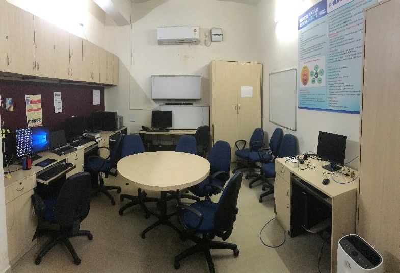
Gallery
Faculty

Contact Us
Address:
Google Map: https://goo.gl/maps/EJZ9fsaKfooUxUhy6
Email: radiology.kmc@manipal.edu
Phone Number: 08202922120